ExoSurge® Technology
ExoSurge® Inter-fibrinous Pulsated Gas and Drug Technology (IPG)
Five issued patents, 12 patents and provisionals filed both nationally and internationally

The ExoSurge® Inter-fibrinous Pulsated Gaseous Technology (IPG) uses a sequenced three-step pulsated gas injection followed by injection of an interlesional medicine within every 20-minute ExoSurge treatment. This combination has been proven to cause measurable and permanent reduction of penile fibrosis plaques. Our new pulsated gas and drug treatment technology has resulted in 15 patent filed or granted applications from our team of medical professionals and engineers.
Our Inter-fibrinous Pulsated Gas Technology (IPG) delivers a treatment that is well tolerated, generates no heat or collateral tissue damage, requires no patient downtime or anesthesia, and works without regard to patient skin color.
IPG technology is a novel and distinctive method of drug administration. It has been clinically proven to transform long-known generic intralesional Peyronie's medications into "super drugs" that produce curative results in which Peyronie's fibrosis is broken up and dissolved into the body, just as is expected to occur when the body initiates a fibrotic response in response to injury. The pulsated gas injections open small pathways within the dense fibrous plaques, causing injected medicines to penetrate deeper and with more effectiveness.
Dive deeper into the technology and our plans for the future from the link below.

Our New Medtech Drug Delivery Device

Our clinically proven and patented ExoSurge® technology uses injected and pulsated gases in tandem with intralesional drug injections to cause the permanent removal of fibrous tissue plaques
associated with a Peyronie's Disease diagnosis.
Our years of research and development devoted to unlocking the epidemiology of Peyronie's plays a key role in our technology's success.
We are science-driven and led by executives and medical advisors. Let us show you our plans to revolutionize the treatment of Peyronie’s disease.
Collagentics®
Upon the finalization of our new patent, we will disseminate further information regarding this significant discovery.

Important Breakthroughs from ExoSurge® IPG Technology for Clinicians
1. ExoSurge® Inter-fibrinous Pulsated Gas and Drug Technology (IPG) delivers a treatment that is well tolerated, generates no heat or collateral tissue damage, requires no patient downtime or anesthesia, and works without regard to patient skin color.
2. The methodical IPG process of breaking down and permanently removing penile fibrosis and plaques is a game-changing new tool for Peyronie’s care.
3. ExoSurge® proved effective treating recent acute penile injury cases still in the active phase. Not only did ExoSurge therapy eliminate acute pain within about a week after starting treatments but also healed injured tissue and halted the build up of fibrosis before it became a problem in 100% of cases.
4. The technology proved effective treating diffuse plaque as discovered from duplex Doppler sonogram images (early stage Peyronie’s), completely healing the disease before it became problematic.
5. ExoSurge® paves the way for preventative care in asymptomatic patients who exhibit plaque and fibrosis during pre-screening ultrasound while being evaluated for other urologic disorders such as erectile dysfunction.
6. ExoSurge® treatments can be performed by a urologist or their support staff such as a nurse practitioner, physician’s assist or licensed medical doctor.
7. The device can conveniently be stored in the corner of an exam room between uses. It’s complete size is approximately 48 inches tall by 24 inches wide.

Retrospective Study
Outcomes of Non-Surgical Treatment for Peyronie’s Disease: Retrospective, single clinic, observational cohort study
By:
Steven L. Morganstern, MD
Kenneth J. Carney, MD, PharmD, FACS
Angela Bates, APRN
Abstract
Objectives: The goal of this study is to evaluate the outcomes of a non-surgical treatment for Peyronie’s Disease.
Peyronie’s Disease (PD) is recognized as a difficult disease to treat within the medical community. The basis for difficulty may stem from the controversy in agreement on the etiology of PD (Nehra, et al., 2015). Most treatments to date are centered around treating the symptoms caused by the disease or for discouraging the initiation of fibrosis (Fallo & Sarnacchiaro, 2019) (Gonzalez-Cadavid & Rajfer, 2010). At this time there is only one treatment approved by the FDA (U.S. Food and Drug Administration) for non-surgical intervention of PD, clostridium collagenase histolyticum (CCH). This treatment can be effective however due to stringent qualifications for use, many patients suffering with PD are excluded from this treatment (Nehra, et al., 2015). Recommendations for treatment from the American Urological Association (AUA) include several options for medication usage with varying degrees of expected success, other than CCH. The evidence listed for these medications is rated as Grade C. This level of reliability in the research shows an expectation toward change in the confidence level with further research (Nehra, et al., 2015).
Materials and Methods: Of the 101 subjects reviewed, 31 patients were excluded due to a lack of continuity in record keeping leaving 70 patients who were actively in treatment between the dates of January 1, 2018 and March 6, 2020. Peyronie’s treatment using Exosurge follows current guidelines provided by the AUA for Peyronie’s Disease (Nehra, et al., 2015). No patients were excluded for past medical history, fibrotic condition of calcification, degree of initial curve or previous treatments.
Results: Changes in presentation after treatment resulted in 84% of patients with improvement. Of these patients, there was a 58% average reduction in curvature. Fourteen percent showed no change and only one patient worsened after treatments began.
The average plaque reduction was found to be 79%. Patients who had an initial curve showed an average plaque reduction of 88%. An incidental finding revealed that only 24% of patients with a curve greater than or equal to 30 degrees measured an acceptable erection grade of 3.5 or 4.0 at baseline. After treatments, this number increased to 42%.
Conclusion: Not only do these results show an impressive reduction in plaque size and consistency, but a notable difference was seen in the degree of curve and erectile function. This author feels there is enough evidence presented within this review to proceed to clinical trials for the use of ExoSurge as a foundational, non-surgical treatment for Peyronie’s Disease.
Key Points:
· 84% of patients showed improvement in their curve.
· 58% average reduction in curve.
· 79% overall reduction in plaque.
· 55% of patients with an initial curve greater than 30 degrees achieved an erection grade of at least 3.5 after treatment. This is an increase of 31%.
Outcome of Non-Surgical Treatment for Peyronie’s Disease: Retrospective, single clinic, observational cohort study
Introduction
Peyronie’s Disease (PD) has been defined as a disease of the tunica albuginea in which fibrosis and plaque formation alter the physiology of the corpus cavernosum. The presentation of this plaque may cause a singular or a combination of changes such as erectile dysfunction, pain, curvature, reduction in girth and length as well as a “wasting effect” of the penis (Hussein, Alwaal, & Lue, 2015). During a systematic review performed by the AUA (Nehra, et al., 2015), a definition of PD was constructed as an abnormality characterized by fibrosis. Further, it was reported that PD may include pain, erectile dysfunction, or a deformity. Although symptoms are included in the definition of PD, it should be recognized that the base definition of PD is fibrosis.
Treatment for PD is controversial as most treatment modalities have minimal success and few show a continued progress toward resolution. Many known treatments only address the symptoms that present with PD and do not look to the cause. The AUA recommends that clinicians should only offer treatment if the clinician is experienced to treat the condition (Nehra, et al., 2015). The purpose of this study was to review the outcomes of PD patients undergoing the non-surgical treatment, ExoSurge at a single clinic, Morganstern Urology Clinic (The Clinic) between the dates of January 1, 2018, and March 6, 2020. No patients were excluded based on past medical history or type of treatment received in the clinic.
2 Method
2.1 Eligibility criteria for participants
A database search using The Clinic’s Electronic Health Records (EHR) system was completed extrapolating patient medical charts with the diagnosis of PD and undergoing treatment between the dates of January 1, 2018, and March 6, 2020. Patient charts were included when they were found to have identifiable and measurable plaque with no less than two duplex doppler ultrasound diagnostic tests within that timeframe providing a pre-treatment ultrasound and a post or intra-treatment ultrasound for comparison. Of these patients, 101 were found to be consistent in receiving treatments during this timeframe. Those excluded were patients who had not been seen in the clinic within three months or more prior to the post or intra-treatment ultrasound tests and considered inconsistent with treatment.
Upon further review it was determined that within this patient sampling of 101, some pre-treatment recordings of the duplex doppler ultrasound diagnostic test was not complete and/or not comparable in recording to the post and/or intra-treatment ultrasound and therefor were not considered a good comparison. These patient charts were removed from the study leaving a total of 70 patient charts for comparison. All duplex doppler ultrasound diagnostic testing during the listed dates was completed by an independent, outside source on site at The Clinic.
2.2 Past Medical History
Past medical history (PMH) for each patient was recorded only. No participants were excluded based on PMH. Past medical history was broken down into these categories: Cardiac, Diabetes Mellitus (DM), Cancer (CA), Cerebrovascular Accident (CVA) and Low Testosterone (Low T). The heading of cardiac included patients with diagnosis and having been medically treated for hypertension (HTN), hypercholesterolemia, arrhythmia, congestive heart failure (CHF) or had a history of myocardial infarction (MI). Patients were included in the diabetes category if they were found to have a history of being treated medically or were currently being treated for diabetes mellitus (DM).
2.3 Study Design
This was a single clinic, descriptive study conducted in review of clinical treatments by Morganstern Urology Clinic including patients undergoing treatment for Peyronie’s Disease between January 1, 2018, and March 6, 2020. Patient identification information was removed to maintain HIPAA Compliance.
Data retrieved from each duplex doppler ultrasound included degree of curve, identification of venous leak, identification of arterial insufficiency, erection grade, size of plaque, and consistency of plaque as either calcified or non-calcified.
2.4 Treatment Description
ExoSurge treatment includes a gas combined with an intralesional injection of no less than 1 mL of the calcium channel blocker verapamil. Other supportive treatments were used based on each patient’s individual presentation, but ExoSurge and Verapamil were administered to every patient in this study. Supportive treatments may have included straight stretching devices, vacuum devices, oral medications, intralesional medications and/or over the counter supplements.
2.5 Data Review & Analysis
A beginning baseline duplex doppler ultrasound analysis that included up to three measurable pieces of plaque was compared to a follow up duplex doppler ultrasound for measurable changes. Data retrieved from both the initial and final duplex doppler ultrasounds included findings of plaque. Plaque size was recorded in a 3-dimensional format in millimeters. Consistency of plaque was recorded as calcified or non-calcified. Penile curvature was identified in degree of curvature, which was then divided into two classes: curvature greater than or equal to 30 degrees and a curvature less than 30 degrees.
Vascular integrity was recorded by review of arterial flow and/or venous leak. A simple A to B Ratio was used to determine level of integrity. The average value of Peak Systolic Velocity (PSV) was recorded per sides left and right. The average value of End-Diastolic Velocity (EDV) was also recorded by sides left and right. These results were then divided into Healthy, Moderate or Severe and noted as left and right. An A/B ratio greater than 20 was labeled as healthy, between 7 and 19 as moderate and less than 7 as severe. For final review only results shown to have Moderate or Healthy in both the left and right side were recorded as “unhealthy”.
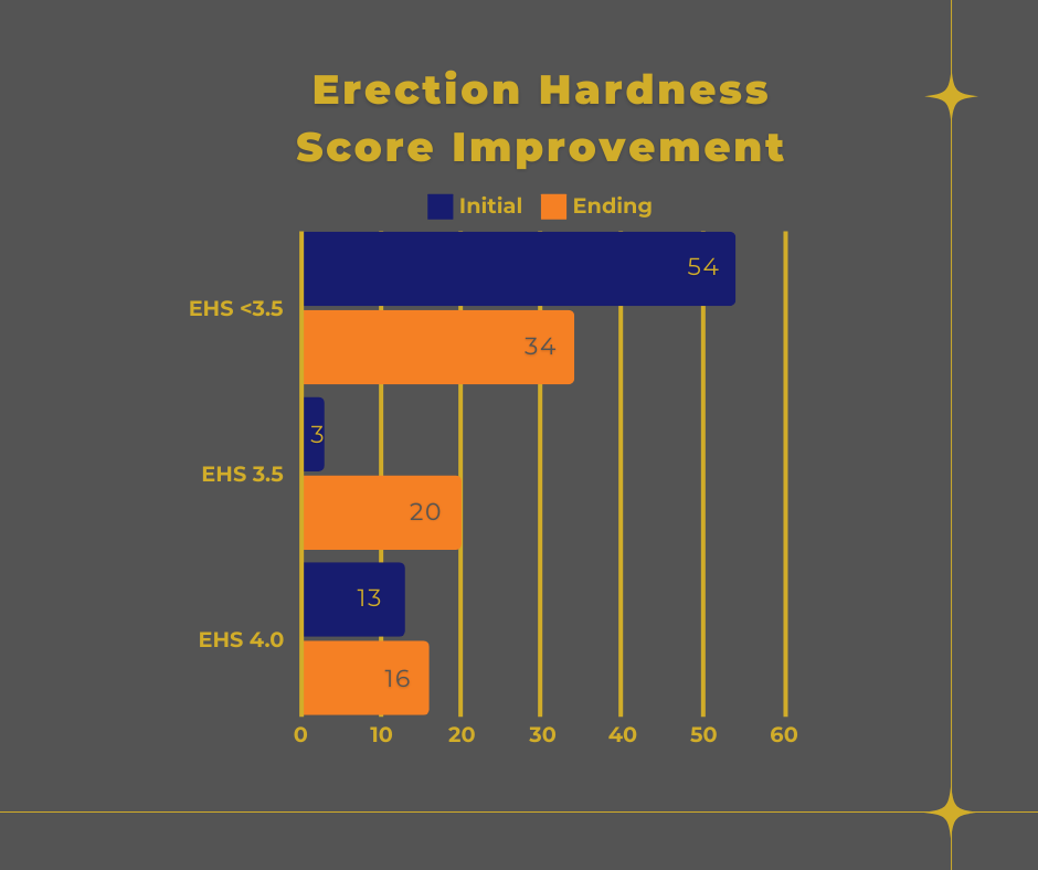
Erection grade was recorded on a scale of 1 to 4 with increments of 0.5 based on the Erection Hardness Score (EHS).
Figure 1 EHS (Goldstein, et al., 2008)
2.6 Statistical Analysis
Of the total 70 patients reviewed, 50 were found to have a curve not considered as natural and developing later in life. All 50 patients were evaluated with color duplex doppler ultrasound for the following variables: penile plaque volume, calcified vs. non-calcified plaque, degree of penile curvature, and aspects of erectile function. Existing health conditions were noted but have not been used to score or further rate any changes in the level of disease for this review.
Plaque volume was measured, in mm3. To calculate the volume of the penile plaque, we measured three dimensions of each piece to determine the volume; V = length ×width × depth. Up to three separate pieces of plaque were recorded when present.
Degree of penile curvature was recorded during the initial and follow up duplex doppler ultrasound by use of a protractor. In many cases, but not all, pictures were provided with protractor showing measurement of current curve. Value of curve was accepted with or without confirmation picture.
Each duplex doppler ultrasound included an intra-cavernosal injection of 10 mcg alprostadil (Nehra, et al., 2015) administered prior to any measurements (Cavallini, Scroppo, & Zucchi, 2016). PSV was recorded every five minutes for a total of thirty minutes on both the left and right sides. For purposes of evaluation, PSV readings less than 30 were considered deficient (Gomez Varela, Mateos Yeguas, Rodriguez, & Duran Vila, 2020) and recorded as “arterial insufficiency”. EDV was also recorded in the same increments. It is commonly recognized that the EDV must be low to offer an effective veno-occlusive mechanism (Cavallini, Scroppo, & Zucchi, 2016). For evaluation, we considered readings >4 cm/sec as insufficient and this is labeled as “venous leak”. A simple ratio calculation of PSV / EDV provided the ratio between the two ratings.
3 Results
3.1 ExoSurge treatment changes in Plaque size
3.1A Plaque Area One Condition:
For the baseline duplex Doppler ultrasonography, the total number of pieces counted were 66. Of these, 52 were non-calcified and 14 were calcified. On the follow up duplex Doppler ultrasonography, the total number was 58. Of these, 48 were non-calcified and 10 were calcified. The reduction in the number of calcified plaque pieces was 28.6%.
3.1B Plaque Area One Measurement:
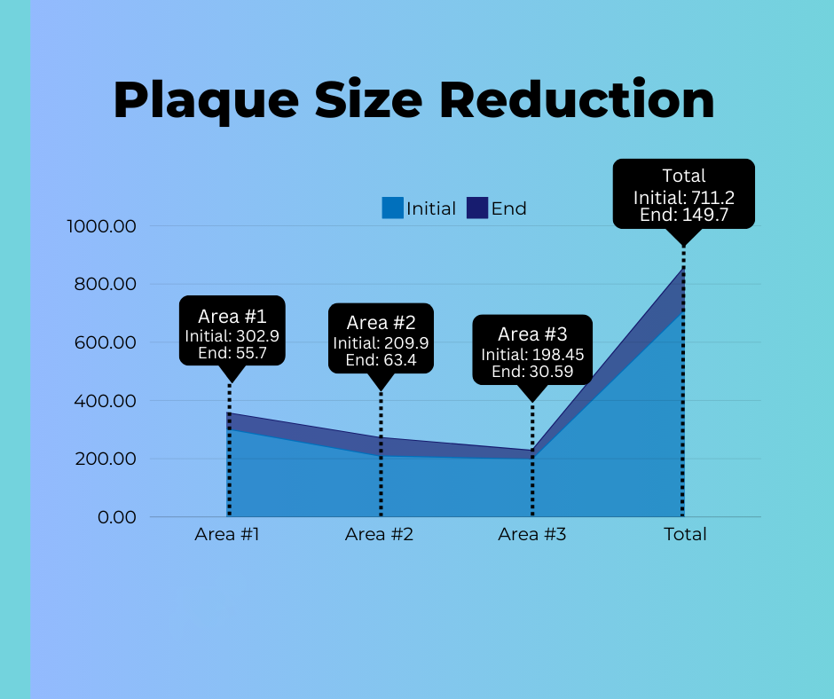
The total plaque size measured with the baseline duplex Doppler ultrasonography in Area One was 302.9 mm3 and the follow up duplex Doppler tally in Area One was 55.7 mm3, representing an 81.5% reduction in penile plaque for the 70 patients.
3.1C Plaque Area Two Condition:
For the baseline duplex Doppler ultrasonography, the total number of pieces counted in plaque area Number Two were 42 with the follow up study showing a total of 40. Of the original 42 pieces, 28 were non-calcified and 14 were calcified. On the follow up duplex Doppler ultrasonography analysis for plaque in Area Two, 32 were non-calcified and 8 were calcified, a 43% decrease in calcified plaque.
3.1D Plaque Area Two Measurement:
The average total size as measured from the baseline duplex Doppler for area two was 209.9 mm3 and the follow up results measured 63.4 mm3, representing a 70% reduction in penile plaque.
3.1E Plaque Area Three Condition:
For the baseline duplex Doppler ultrasonography, the total number of pieces counted in plaque Area Three were 22. The follow up duplex Doppler ultrasonography showed a total of 23 plaque pieces. Of the original 22 pieces, 15 were non-calcified and 7 were calcified. Data derived from the follow up duplex Doppler ultrasonography indicated 18 plaque fragments were non-calcified and 5 were calcified, representing a 28.6% improvement in the reduction of calcified plaque.
3.1F Plaque Area Three Measurement:
The total size as measured with the baseline duplex Doppler ultrasonography study for plaque in area three was 198.45 mm3 and the follow up duplex Doppler ultrasonography was 30.59 mm3, representing an improvement in the reduction of penile plaque of 84.6%.
Figure 2 Plaque Size Reduction
3.1G Combined Areas Condition:
The initial baseline number of calcified plaque pieces derived from duplex Doppler ultrasonography among all 70 patients in this study was 35. The follow up duplex doppler study showed the total number of calcified plaque pieces after treatment was 23. This shows a 34.3% reduction in calcified plaque pieces.
The non-calcified numbers increased from an initial count of 95 to the follow up count of 98. This is an expected finding as the calcified plaque will change to non-calcified before it is no longer measurable.
3.1 H Combined Areas Measurement:
The initial baseline plaque size total derived from duplex Doppler ultrasonography among all 70 patients in this study was 711.2 mm3. The follow up duplex Doppler ultrasonography was 149.7 mm3. The combined measurable penile plaque reduction for all areas was 79.0%.
Peyronie's Disease Study - Plaque Size Reduction
Figure 2 Penile Plaque Size Reduction
3.2 ExoSurge treatment change in curve
A comparison was made between initial duplex doppler ultrasound and final/intra-treatment ultrasound as some patients were still undergoing treatments. Of the initial 50 patients with a curve, 36 of those patients also had a venous leak. Of the 50 patients with a curve, 17 of them had a curve less than 30 degrees with 12 of those with a venous leak. Thirty-three of the 50 had a curve greater than 30 degrees, of which 24 had a venous leak. The final study review of the 50 pts who were initially found to have a curve, 8 of those patients had no curve at all. Of the original 33 with a curve greater than 30 degrees, this number decreased to only 20 patients. On the initial study only 17 patients had a curve less than 30 degrees and this increased to 22. Therefore, 8 patients completely corrected to no longer have a curve and 5 had reduced significantly, one patient's curve increased by 8 degrees. A 58% reduction in curve was noted for the entire group who showed a decrease in the degree of curve. Fourteen percent of patients showed no improvements with the curve.
During the time reviewed, we found only 24 patients still receiving therapy at the final date for review. The individual average improvement for these patients at the end date was 60%. Of the 26 patients no longer participating in treatment, there was an individual average
Peyronie's Disease Study - Average Improvement
Figure 3 Improvements Average Per Patient
improvement of 55% to their curvature. This change is congruent with our observations showing the largest change in curvature realized toward the end of therapy.
3.3 ExoSurge treatment changes in erectile dysfunction
Erection Hardness Score Improvement
Figure 4 EHS Patient Improvement
This study was a Peyronie's disease/plaque study and shows reduction in volume of plaque however, the results may be easier to see when reviewing the changes recognized in the degree of curve. Of the total 70 patients reviewed, 16 were initially found to show an erection grade of 3.5 or higher. The data reviewed is the measured erections grade found on the first and last vascular study to allow for comparison. Initially, of the total 70 patients, 13 patients were evaluated at an erection grade of 4, after treatments there was a total of 16 patients. Initially only 3 pts were graded at a 3.5 but there was a total of 20 patients on the re-evaluation. It could be that those 3 patients who were graded at a 3.5 moved up to a 4 and the 20 came from patients who initially were found with erections grades at a 3 or lower. Therefore, to begin with only 16 patients had erection grades of 3.5 or 4 out of the original 70 participants but afterward 36 patients showed a grade of 3.5 or 4.
4 Discussion
Although this study was to review the success or failure of a new, non-surgical technique for correction of Peyronie’s Disease the data obtained has value in treatments for erectile dysfunction related to venous leak and arterial insufficiency. Further review is necessary to identify the variables and obtain a more in-depth presentation toward future treatment recommendations for ED. Within the cohort information collected for this review, there is sufficient data for other conclusions related to abnormalities of the penile shaft.
Many of the tools used during the initial and subsequent duplex doppler ultrasound studies were standardized to the industry however the A/B ratio used for review of PSV to EDV was not. It appears to be sufficient to identify venous leak levels in conjunction with arterial flow for this data review, however the Resistive Index (RI) is more common practice and should be evaluated for future use. Although the A/B ratio is used in The Clinic for determining different treatment options, it does not seem to directly correlate with erection grade.
Surprisingly, there appeared to be a connection with patients who initially showed a venous leak alone or in combination with arterial insufficiency and their original erection score. This data should be reviewed for analysis of vaso-occlusive erectile dysfunction.
Based on our findings, it would be of interest to review and compare each patients PMH along with their level of treatment to see to what degree these disease processes affect the treatment of Peyronie’s Disease (Herrera, Henke, & Bitterman, 2018) (Ming, Ma, Xhang, Guo, & Yuan, 2020) and not just the disease itself.
5 Conclusion
The review of data has shown significant improvements with patients undergoing ExoSurge treatments and Verapamil. This data proves this to be a successful foundational treatment for Peyronie’s Disease in patients of all conditions and medical histories. Not only did these results show an impressive reduction in plaque size and consistency, but a notable difference was also seen in degree of curve as well as erectile function. Current guidelines from the AUA for the treatment of Peyronie’s Disease include several options for treatment and a category named “Other Treatments”. Also included are recommendations for surgical intervention if a patient presents with erectile dysfunction along with PD (Nehra, et al., 2015). The results indicate that not only is there is enough evidence presented to proceed to clinical trials for the use of ExoSurge as a, non-surgical treatment for Peyronie’s Disease but also shows that improvements can be made in PD patients who experience ED as well. We should not be going directly to surgical intervention for patients with ED. This treatment continues to follow current guidelines provided by the AUA while avoiding recommended irreversible surgical interventions such as penile prosthesis or grafting.
5 Authors Notes/Conflicts of Interest
Authors are current members of the care team at Morganstern Urology and active during treatment administration, data assimilation and extrapolation.
References
Brock, G., Hsu, G., Nunes, L., von Heyden, B., & Lue, T. (1997, Jan). The anatomy of the tunica albuginea in the normal penis and Peyronie's disease. Jurnal of Urology, 157(1), 276-81.
Cavallini, G., Scroppo, F. I., & Zucchi, A. (2016). Peak systolic velocity thresholds of cavernosal penile arteries in patients with and without risk factors for arterial erectile deficiency. Andrology. doi: https://doi.org/10.1111/andr.12242
Connolly, J. A., Borirakchanyavat, S., & Lue, T. F. (1996). Ultrasound evaluation of the penis for assessment of impotence. Journal of Clinical Ultrasound. doi:https://doi.org/10.1002/(SICI)1097-0096(199610)24:8<481::AID-JCU8>3.0.CO;2-G
Fallo, L., & Sarnacchiaro, P. (2019, April). Ten-year experience with multimodal treatment for acute phase Peyronie's disease: A real life clinical report Ten years of experience with the multimodal treatment of the acute phase of Peyronie's disease: real-life medical report. Spanish Urological Records (English Edition), Online 1-3. doi:10.1016//J.acuroe.2019.03.007
Goldstein, I., Mulhall, J. P., Bushmakin, A. G., Cappelleri, J. C., Hvidsten, K., & Symonds, T. (2008, Oct 8). The Erection Hardness Score and Its Relationship to Successful Sexual Intercourse. (10, Ed.) The Journal of Sexual Medicine, 2374-2380. doi:10.1111/j.1743-6109.2008.00910.x
Gomez Varela, C., Mateos Yeguas, L. A., Rodriguez, I. C., & Duran Vila, M. D. (2020). Penile Doppler Ultrasound for Erectile Dysfunction: Technique and Interpretation. American Journal of Roentgenology, 214(5), 1112-1121.
Gonzalez-Cadavid, N. F., & Rajfer, J. (2010). treatment of Peyronie's disease with PDE5 inhibitors: an antifibrotic strategy. Nat Rev Urol, 7, 215-221.
Herrera, J., Henke, C. A., & Bitterman, P. B. (2018, 01 2). Extracellular matrix as a driver of progressive fibrosis. J Cli Invest, 128(1), 45-53. doi:10.1172/JCI93557
Hussein, A. A., Alwaal, A., & Lue, T. F. (2015). All about Peyronie's disease. Asian Journal of Urology, 2, 70-78.
Lue, T. F., Kricak, H., Marich, K. W., & Tanagho, E. A. (1985). Vasculogenic impotence evaluated by high-resolution ultrasonography and pulsed Doppler spectrum analysis. Radiology, 155(3). doi:https://doi.org/10.1148/radiology.155.3.3890009
Ming, L., Ma, X., Xhang, X.-L., Guo, L.-Q., & Yuan, M.-Z. (2020). Significance of blood lipid parameters as effective markers for arteriogenic erectile dysfunction. Andrology, 8, 1086-1094. doi:doi.com/10.1111/andr.12776
Nehra, A., Alterowitz, R., Culkin, D., Faraday, M. M., Hakim, L. S., Heidelbaugh, J. J., . . . Burnett, A. L. (2015). Peyronie's Disease: AUA Guideline. Journal of Urology, 194, 745-753. doi:10.1016
Padma-Nathan, H., Stecher, V. J., Sweeney, M., Orazem, J., Tseng, L.-J., & Deriesthal, H. (2003). Minimal time to successful intercourse after sildenafil citrate: results of a randomized, double-blind, placebo-controlled trial. Urology, 62(3), 400-403. doi:doi: 10.1016/s0090-4295(03)00567-3
Prajapati, D., Rampal, K., Ali, I., Rangera, M., Chaurisia, S., & Prajapati, J. M. (2016). Penile fracture and its managment. International Surgery Journal, 3(4), 1714-1717. doi:10.18203/2349-2902.isj20163552
Wynn, T. A. (2009, Jun). Cellular and molecular mechanisms of fibrosis. J Pathol, 214(2), 199-210. doi:10.1002/path.2277
Table of Figures
Figure 1 EHS (Goldstein, et al., 2008)
Figure 2 Plaque Size Reduction
Figure 3 Improvements Average Per Patient
Figure 4 EHS Patient Improvement
Existing Peyronie’s Disease Treatments
To date, therapies for treating Peyronie’s disease only vie to improve symptoms of Peyronie’s Disease patients. None heal the underlying basis for the disease. None are permanent. None are effective with every case. Thus, at best, existing treatments offer some patients a temporary reprieve from the worst symptoms of Peyronie’s Disease.
ExoSurge® is the world’s first clinically-proven treatment to conservatively and permanently remove plaques and fibrosis associated with a Peyronie’s diagnosis.
Peyronie’s Disease Redefined
We have redefined Peyronie’s disease based upon our insights from curative outcomes. As such, we define Peyronie's as the build up of fibrous tissue plaques located within the soft tissue of the penis and/or thickening of the tunica albuginea. This build-up of these non-cancerous tumorous masses can result in a myriad of negative functional and misshaping of a penis including curvature when erect, indentions to the erect shaft, lost length, loss of adequate blood flow to form an erection, hour glass shaping, etc.
Many of these conditions worsen with time. The body often sends additional fibrosis to the affected area in an attempt to help heal the condition.
Furthermore, any calcification in the bloodstream eventually accumulates on top of pre-existing fibrotic masses, resulting in a much more dense and problematic case known as "calcified plaque".
Current FDA-Cleared Peyronie’s Disease Treatments
Approvals and clearances by the FDA for Peyronie’s treatments to date have only evaluated changes in potential symptoms that can be caused by the build of plaques and fibrosis within the penis, such as the degree of erect curvature. Significant erect curvature is a common symptom of a Peyronie’s diagnosis affecting about 70% of patients. Based upon our research, the degree of erect curvature is tied to the exact size and location of the fibrous tissue plaques.
All of the negative side effects of Peyronie’s Disease are tied to the build up of penile plaques, in the soft tissue of penis, which is the true definition of Peyronie’s disease.
ExoSurge® is the world’s first clinically-proven therapeutic to measurably and permanently remove fibrous tissue plaques from the penis.
Collagenase clostridium histolyticum injections
Known under the brand name Xiaflex®, collagenase clostridium histolyticum is an enzyme that purports to breaks down the collagen scar tissue found in the plaques that cause PD. Xiaflex® is the only injectable drug approved by the U.S. Food and Drug Administration for the treatment of PD, and is administered directly into the plaques on the penis, via injection, following local anesthesia.
Collagenase clostridium histolyticum is very expensive, but it’s covered by most health insurance plans, and Endo Pharmaceuticals sometimes provides a co-pay assistance.
Treatment typically starts with two injections, given 1 to 3 days apart, followed by 6 weeks of penile stretching (see below). This cycle is repeated three more times for a total of eight injections.
Though sometimes effective, CCH injections won’t completely resolve penis curvature from PD, and they won’t restore lost length or girth. In addition, in rare instances, injectable treatments can cause rupture of the penile tissues that will require surgical repair.
Verapamil
Verapamil is a generic antihypertensive drug and calcium channel blocker, which is most commonly used for treating high blood pressure. Research suggests that calcium channel blockers increase the activity of collagenase, an enzyme that helps breaks down the excess collagen that makes up PD plaques.
Verapamil injections are usually covered by health insurance plans if performed by a urologist. It’s administered as an injection every two weeks for 12 weeks, for a total of six injections. Injections are administered directly into the affected area of the penis, under local anesthesia.
Improvement from Verapamil injections is historically minimal.
Restorex® Penile Traction Therapy (PTT)
There are countless penile traction devices on the market, However Retorex® is the only version that's FDA-cleared.
We love Restorex® and we now recommend it to most of our patients getting ExoSurge® treatment. It's an exceptionally well-engineered device compared to prior iterations for penile traction. That advanced design and components are reflected in its $500 price but it's money well spent.
The most frequently encountered adverse side effects of penile traction are mild pain, skin irritation, bruising and skin discoloration, and itching.
During ExoSurge's first retrospective study, where our patients with erect curvature saw a 60% improvement, Restorex® was not used.
We believe our reported curvature results would have been significantly better if patients had used Restorex penile traction device at that time.
Peyronie’s Surgery
Surgery remains the gold standard for Peyronie’s care according to the AUA's standards of care and it comes in two basic pathways: "incision and graft" and "plication".
Incision and graft is usually only indicated for the most severe cases. However, the incidence of recurrence and erectile dysfunction is significant, and some individuals may experience irreversible loss of penile sensation.
A retrospective study published by Florian Wimpissinger, MD, of the department of urology at Rudolfstiftung Hospital in Vienna, Austria in 2012[2] regarding the long-term outcomes associated with PD surgery showed a notable recurrence of Peyronie’s symptoms over time.
After a mean of 9.3 years, the recurrence of postoperative curvature had increased from 0% after a mean of 38 months (18–73 months) to 23.7%, post-op erectile dysfunction increased from 3.0% to 39.5%, penile shortening from 0.0% to 65.8%, and impairment of penile sensitivity from 3% to 31.4%.
“We saw that results tend to worsen with time,” said Dr. Wimpissinger, who presented the study results at the 2011 European Association of Urology annual congress in Vienna.
Plication surgery has the most reliable outcome and the least chance of ED, but it is unacceptable for many patients due to the "loss" of penile length. The post-surgical penile length is usually around where the erect curvature begins along the penis shaft. Very few men are comfortable losing 1-3 inches of erect length regardless of circumstances.
Non-Traditional Peyronie’s Disease Treatments
We always explore and stay abreast of new ideas and innovations for Peyronie's care.
We are publishing a new report soon to explore non-traditional therapies and their possible role in Peyronie's care for the future.

ExoSurge® Inter-fibrinous Pulsated Gas and Drug Technology (IPG)
The ExoSurge® Inter-fibrinous Pulsated Gas and Drug Technology (IPG) uses a sequenced five-step, single visit, 20-minute treatment process that results in measurable permanent reduction of penile fibrosis with repeated applications.
Our inter-fibrinous gaseous injections are a novel and distinctive method of drug administration as they're able to penetrate plaques and open pathways to medicines. They have been clinically prove to transform long-known generic intralesional Peyronie's medications into "super drugs" that produce curative results in which Peyronie's fibrosis is broken up and dissolved into the body, just as is expected to occur when the body initiates a fibrotic response in response to injury.
The combination of new pulsated gas technology and intralesional drugshas resulted in 15 patent filed or granted applications from our team of medical professionals and engineers.
Our accomplishments in studying the epidemiology of Peyronie's disease are critical to our success.
ExoSurge® researchers have identified six "Peyronie's Accelerators" that not only speed up the onset of Peyronie's symptoms in otherwise healthy patients, but also impair treatment efficacy if left unmanaged during care.
Calcified Plaque: Before & After Sonogram Images
Following are examples of "before and after" duplex sonogram images of ExoSurge® Peyronie's patients with calcified plaques. The reduction of calcified plaques is readily apparent, even to an inexperienced layperson. As far as we are aware, these photos are the first sonographic evidence of a visible reduction in calcified Peyronie's plaque from conservative therapy in the history of urology.


Before and After Peyronie's Treatment Photos
Have you ever Googled "before and after Peyronie's treatment photos"? The only images you'll find are gut wrenching photos taken midway through plication or "incision & graft" Peyronie's surgery.
Following are baseline and follow up images of Peyronie's patients following treatments with ExoSurge®. We hope to share many more soon. Unfortunately, patients must grant us permission to release these photographs, and few are willing to do that.


ExoSurge® is the world's first clinically-proven treatment to conservatively and permanently remove plaques and fibrosis associated with a Peyronie's diagnosis.
ExoSurge® Technology: Refinements and Observations
Between the period of the invention of our first successful Peyronie's treatment technology that achieved measurable improvement with plaque reduction confirmed through our Proof of Study Observations in 2014 to the completion of our 2021 Retrospective Study, we treated hundreds of patients suffering from Peyronie's disease at our clinic in Atlanta with our evolving therapeutic and began to make important observations regarding what the underlying cause and epidemiology of Peyronie's Disease.
When this key pillar was finalized, we became focused upon refinement and improvement of our technology, including:
• Refining our "working-model-theory" regarding the underlying etiology of Peyronie's
• Further study in the nature of penile plaques and fibrosis within varied disease cases
• Patient health factors as a part of a Peyronie's case and diagnosis
• Variations in the most effective means to treat the wide variety of Peyronie's plaques
• Developing weighted scoring for patient's existing health dynamics and their impact on a Peyronie's diagnosis and permanent plaque removal
Continued refinements from case observations to each of these pillars resulted in continued improvement of treatment outcomes. We were late evolving to the use of baseline and followup Duplex Doppler ultrasonography as our measuring stick for patient improvement. We now embrace this "before and after" comparison as the gold standard for the evaluation of Peyronie's treatment
Following is a summation of our most important Breakthrough Observations with Peyronie's Disease made between 2014 and 2021.
To learn more about our diagnostics and Peyronie's treatments with ExoSurge technology right away, please visit our clinical website and schedule a "one on one" phone consultation or a visit to our clinic in the Buckhead area of Atlanta, Georgia.
Stay Up To Date
If you are a Peyronie's patient who has interest in participating in one of our upcoming clinical trials around the United States, please click here
If you are a urologic clinician who has a potential interest with being involved in our upcoming prospective clinical trials, please click here
If you would like to stay abreast of our progress during clinical trials and in the future, please click here:
Sign up to follow our story and to watch us grow!